Protection of tissue physicochemical properties using polyfunctional crosslinkers [full article]
Young-Gyun Park, Chang Ho Sohn, Ritchie Chen, Margaret McCue, Gabrielle T. Drummond, Taeyun Ku, Dae Hee Yun, Nicholas B. Evans, Hayeon Caitlyn Oak, Wendy Trieu, Heejin Choi, Xin Jin, Varoth Lilascharoen, Ji Wang, Matthias C. Truttmann, Helena W. Qi, Hidde L. Ploegh, Todd R. Golub, Shih-Chi Chen, Matthew P. Frosch, Heather J. Kulik, Byung Kook Lim, Kwanghun Chung. Protection of tissue physicochemical properties using polyfunctional crosslinkers, Nature Biotechnology, doi:10.1038/nbt.4281
Introduction
SHIELD is a tissue transformation technology that allows the extraction of multiple biomolecules and multi-scale structures from the same, transparent organ-scale tissue. These unique properties are originated from a polyepoxy chemical. Single polyepoxy has a flexible backbone and multiple epoxide groups that can form intramolecular crosslink with multiple amine groups on the surface of individual biomolecules. Therefore, it can protect tertiary structure of biomolecules and their functions against harsh stressors (e.g., heat, detergents, organic solvents, acidic pH). Polyepoxy can also form crosslinks among multiple endogenous biomolecules to preserve them at their physiological locations and increase the mechanical stability of the tissue-gel.
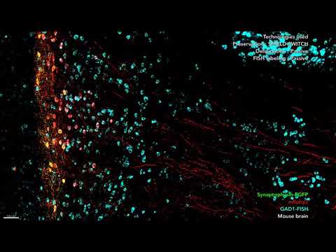
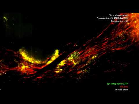
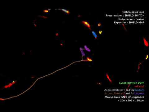
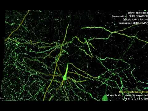
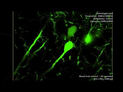
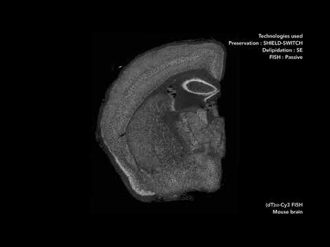
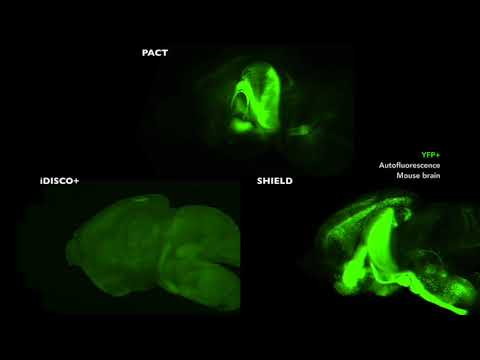
SHIELD preserves protein fluorescence, nucleic acids, proteins, and their probe binding capability (e.g., antigenicity). SHIELD tissue also safeguards native tissue architecture against tissue clearing and de-labeling treatments. Furthermore, combining with MAP (Magnified Analyses of Proteome; Ku et al., Nature Biotechnology, 2016), SHIELD tissue can be expanded 3~4 folds while preserving all key information (protein fluorescence, proteins, nucleic acids, tissue architecture). This allowed us to reconstruct fluorescence-labeled fine subcellular structures from large tissue volume, such as long-range axon fibers and synaptic boutons.

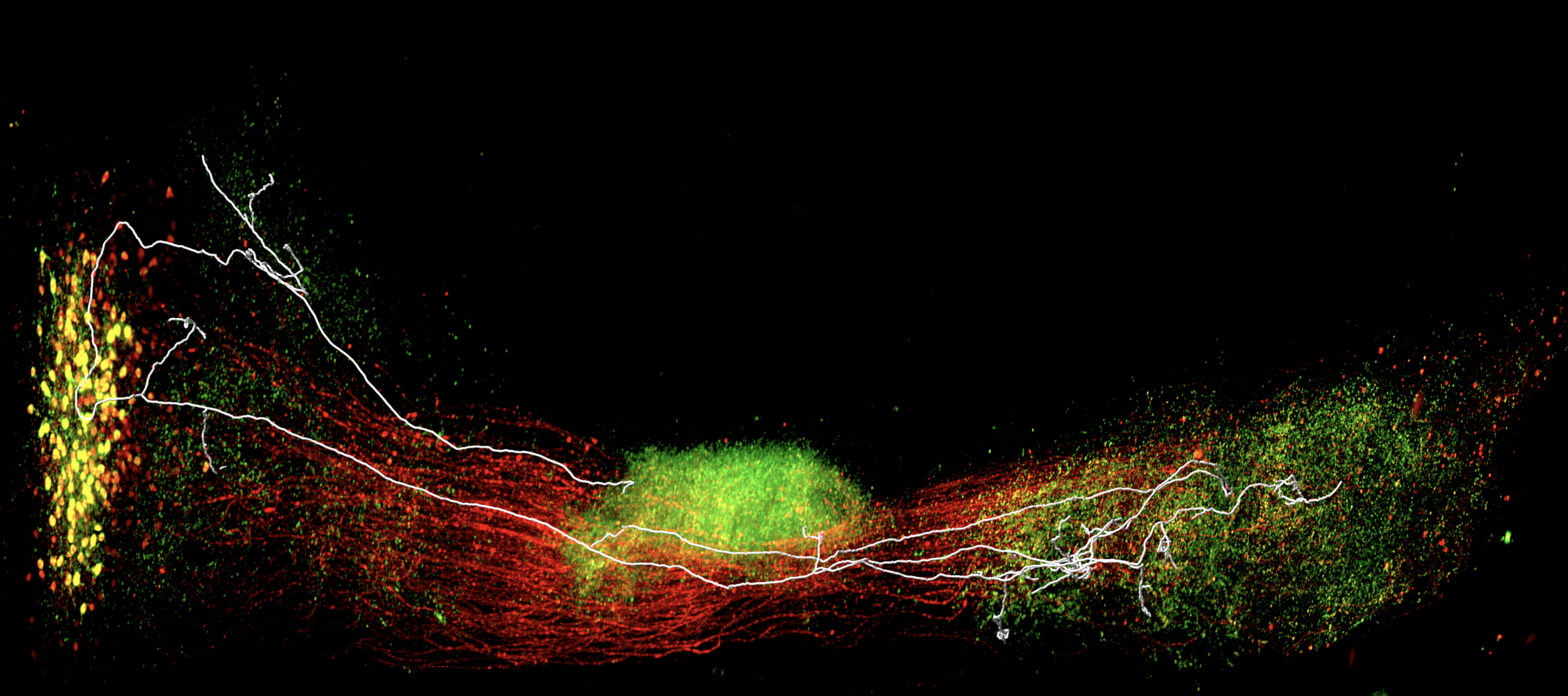
SHIELD enables new approaches in neural circuit mapping. With SHIELD, we could reconstruct an axogram of a single neuron and identify its putative downstream target neurons with their molecular (GAD1 mRNA and Calretinin protein) details.
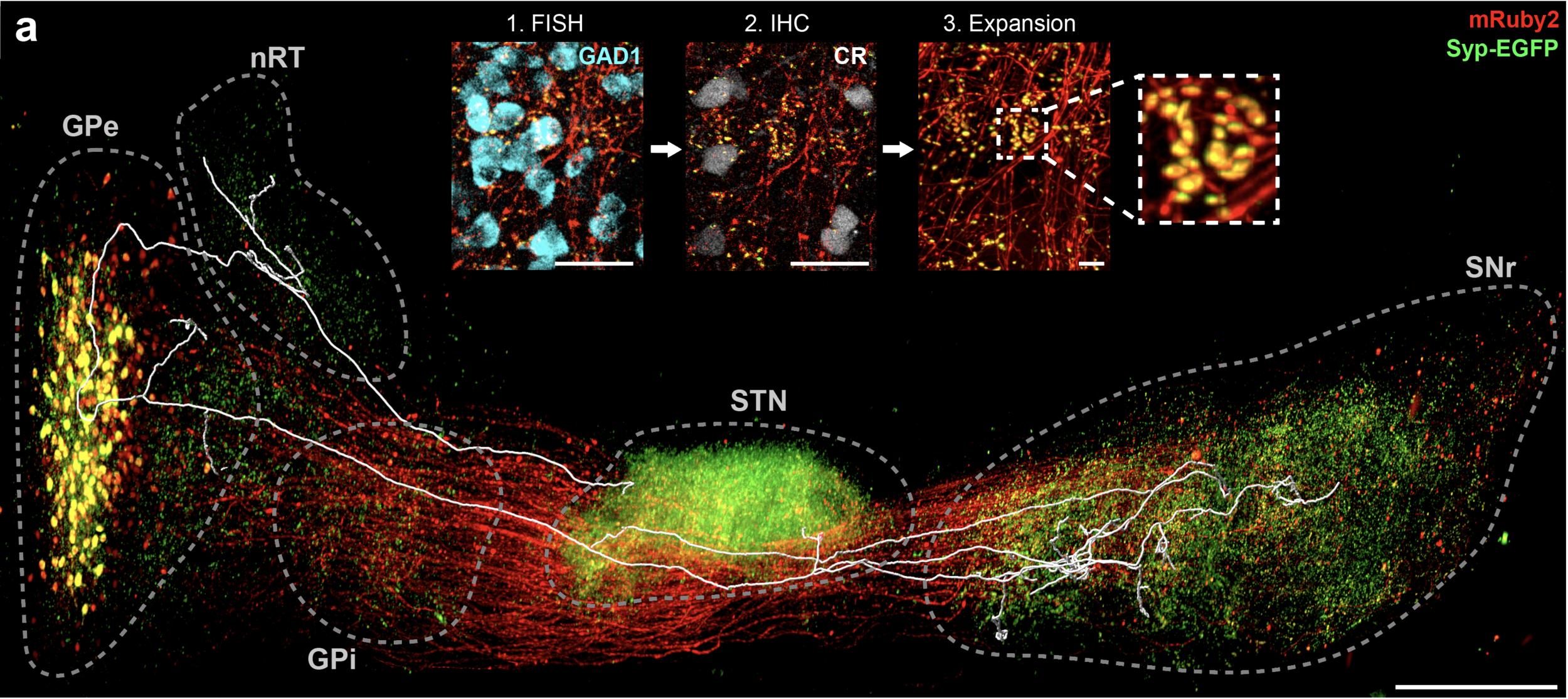

Figure 1. SHIELD enables integrated circuit reconstruction at single cell resolution. (a) 3D rendering of virus-labeled PV+ neurons in GPe with reconstructed axon trace of one of the neuron. Syp-EGFP, Synaptophysin-EGFP. Scale bar : 1 mm. (b) An axogram of the traced neuron and its putative downstream targets. The number of putative axosomatic boutons is marked inside each circle.
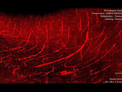
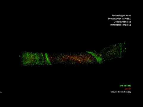
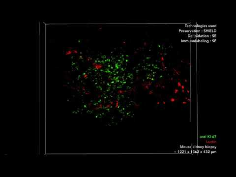
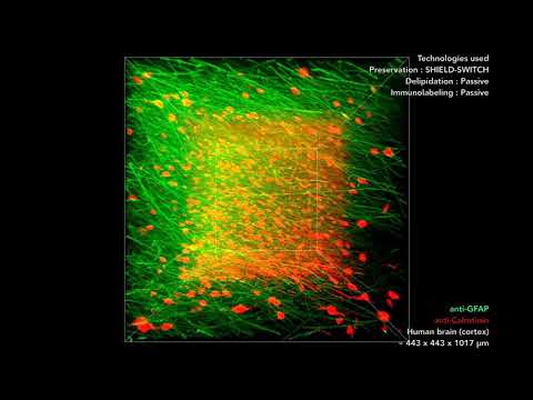
Further, SHIELD is applicable to various types of tissues. For example, SHIELD can transform long-banked human brain tissue into transparent tissue-gel hybrid. SHIELD can be applied for rapid volumetric phenotyping of fresh tissue biopsies. By combining SHIELD with our Stochastic electrotransport (SE) technique, SHIELD allows preserving, clearing, and immunolabeling fresh intact biopsy (1 mm diameter) only within 4 hours.